
SURGICAL PROCEDURES
COUCHING
-
Earliest form of cataract removal
-
Instead of replacing the cataractous lens, it is moved inferiorly to the vitreous cavity, away from the optic axis
INTRACAPSULAR EXTRACTION
-
Introduced in the 19th century
-
Lens was removed, keeping the capsule intact
-
Risk of ocular inflammation and visual loss
EXTRACAPSULAR EXTRACTION
-
Currently used
-
Posterior portion of lens capsule is kept in place
-
IOL replaces the crystalline lens
-
Carried out without phacoemulsification
-
Whole lens nucleus is delivered through an 11mm incision between the cornea and sclera
1967: PHACOEMULSIFICATION
-
Process developed by Charles Kelman
-
Method that degrades the lens using ultrasound waves
-
It is a commonly preferred procedure of cataract extraction
LENS REMOVAL & IOL IMPLANTATION TECHNIQUE:
1). PARACENTESIS
-
A paracentesis is made in the peripheral cornea, distanced away from the main surgical incision, using a 15 degree angle blade
-
The main incision measures around 2.75mm
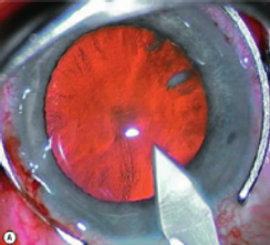
2). CCC
-
Continuous curvilinear capsulorhexis (CCC) process - using forceps to create a tear in part of the anterior lens capsule
-
A flap is created with anterior capsule, and is grasped via forceps to maintain control on the degree of the tear
-
If the flap opening becomes too large, it may allow the IOL to become displaced
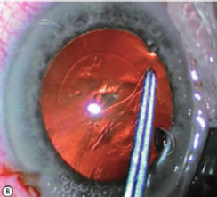
3.) PHACOEMULSIFICATION
-
The next step is to emulsify the lens using a high frequency ultrasonic hand-piece with a titanium or stainless steel tip, known as a ‘phaco probe’
-
The fragments produced as a result of the high energy is aspirated via a pump through the hollow probe tip.
-
The next step is to disassemble the nucleus via a ‘chop’ technique and is aspirated via a vacuum, along with the soft outer lens cortex
4). IOL INSERTION
-
After the removal of the cataractous lens (or to correct a refractive error/ presbyopia), a new intraocular lens is inserted.
-
Typically, a posterior-chamber lens is used, and placed either inside the pre-existing capsular bag, or sometimes in the ciliary sulcus.
-
It is fixed in place via tiny projections known as haptics
-
Mostly, all cataract surgery patients are administered with a local anaesthetic, allowing them to return home the same day.
-
The eye will be considerably recovered in a week, completely recovered in a month
COMPLICATIONS
-
Months/years later some patients posterior capsular opacification (PCO)
-
Here, the capsule becomes opacified behind the IOL and is treated via a YAG laser (Nd: YAG laser capsulotomy), creating a hole in the capsule.
-
This allows for vision to be restored
FURTHER DEVELOPMENTS
-
A femtosecond laser has been introduced, where the patient, under anaesthetic, undergoes a similar procedure where an incision is made in the cornea, capsulotomy is carried out and the cataract is broken up, thus allowing for an IOL to be transplanted
-
This laser technology uses the process of photodissection, where its ultrashort pulses are focused so any collateral and heat- induced damage to surrounding tissues is eliminated